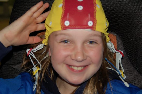
QEEG Brain Mapping

Personalized Neurophysiological Assessment
The last few decades have been very exciting in the field of neuropsychiatry. We no longer have to blindly treat the brain. Advanced neuroimaging tools now allow us to see exactly what is going on with each individual’s brain function. One of these tools is called QEEG, or Quantitative Electroencephalogram.
We now know that dysregulated brain function is the root cause of many cognitive, emotional, and behavioral difficulties. QEEG Brain Mapping offers a sophisticated neuroimaging technology based on a quantitative assessment of the patient’s electroencephalogram. A QEEG Brain Map reveals important information as to how each individual’s brain is functioning. The brain is not simply a chemical organ, but also a neurophysiological organ. This means that it is very important to look at the neuronal activity in the brain. Every brain is unique. Some brains are working too slowly; others are in a state of overarousal. Some brains are marked by inefficient processing speeds while others may struggle with a neuronal firing pattern that is stuck within a particular frequency band rather than flexibly distributing its energy across the frequency spectrum.
The QEEG helps us to know specifically which regions of the brain are involved in the patient’s presenting symptomatology. By comparing the patient’s brain function to a normative database, a personalized medicine approach can be developed to identifying strategies for improving brain function. ADHD has been associated many different Neuroendophenotypic patterns, contraindicating a one-size fits all approach to this condition. The QEEG Brain Map helps us to see why a patient may be struggling with attention or mood and identifies specific frequencies in which the brain’s neurophysiological functioning is imbalanced. This serves to be quite helpful for both neurofeedback and medication treatment directions.
The process starts with a 19-channel recording of the EEG in an Eyes Open and Eyes Closed condition. If the patient is able to minimize artifact for a prolonged period of time, a Cognitive Task condition recording may also be performed. The analysis begins with a visual inspection of the EEG across a wide variety of montages. This is followed by a Fast Fourier Transform Spectral Analysis to determine maximal peaks across 0-30 Hz activity as depicted in the images below.


We are then able to compare readings of Absolute Power, Relative Power, Coherence, Asymmetry, Phase, and Peak Frequency to a normative database to determine the level of statistical significance associated with an individual’s deviation from the norm. This helps to identify target goals for Neurofeedback Treatment Planning, so that the intervention is designed specifically for that person’s unique brain function.

Excess of slow wave Absolute and Relative Power across frontal region bilaterally depicted in the diagram above indicates that this region of the brain is running too slowly in this case of ADHD.
Power Ratios and Relative Power are calculated and compared to norms for the patient’s age to determine in which regions the brain might be imbalanced. The figure below represents an elevated Theta/Beta Power ratio which is often associated with ADHD, Learning Difficulties, and Mood Dysregulation depending upon which region of the brain is involved.

Amplitude Asymmetry, Coherence, and Phase are Connectivity Metrics from the QEEG Brain Map which reveal how efficiently different regions in the brain are communicating with one another. Improved connectivity and synchronous neuronal activity is helpful for efficient information processing.

Low Resolution Electromagnetic Tomography (LORETA) analysis offers specific functional localization analysis as to the 3-dimensional source generators of aberrant brain activity. This noninvasive technology estimates the electric neuronal activity distribution throughout the cortex and subcortical regions, often yielding important information regarding connectivity between regions.

Advanced analysis of the brain’s function under cognitive task (Visual Continuous Performance Test-VCPT) can generate Event Related Potentials (ERPs), which are measures of the brain’s efficiency in sensory and cognitive processing. These can be compared to a normative database to determine areas of inefficiency in the brain’s neuronal activity.
The figure below depicts a normative comparison of ERPs in a case of Traumatic Brain Injury affecting the right hemispheric temporal region. This is an exact neurophysiological correlate to the patient’s presenting difficulties with memory, facial recognition, and comprehension of tone and emotion.

QEEG Brain Mapping offers an exceptional window into the brain’s function. It reveals exactly where the brain is struggling. Clinical Research studies have shown this to be a very accurate, specific, and reliable diagnostic tool. RBH utilizes the QEEG to design individually tailored Neurofeedback Treatment plans.
Call RBH today to inquire if a QEEG Brain Map might be helpful for you or your child!
